Site card
Scanning electron microscope with focused ion beam (FIB-SEM)
Where:
Center for Biomedical Technology
Ubicación:
Centro de Tecnología Biomédica
Typology:
Infraestructura Científica
Manager: Javier de Felipe
Email:
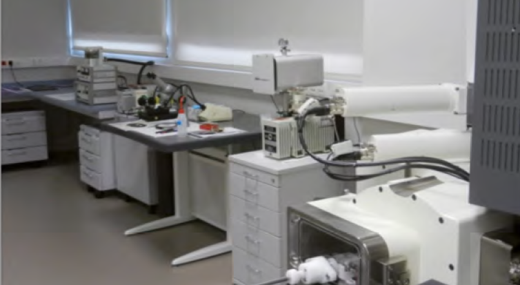
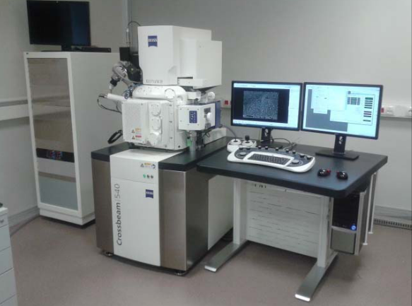
The FIB-SEM equipment consists of a scanning electron microscope (SEM) capable of taking high-resolution images and a focused ion beam (FIB) that can be used to remove successive layers of the sample. The model is ZEISS Crossbeam 540.
Imaging, bioimaging, SEM, surfaces
This equipment is able to performs high-resolution SEM imaging on biological and inorganic materials. In addition, thanks to the FIB, it can take serial images of tissues and reconstruct them in three dimensions.
Serial imaging and 3D reconstruction of biological tissue samples.
The equipment is particularly useful for studying nerve tissue, as it allows for 3D analysis, making it easier to study connectivity at the synaptic level.