Site card
Atomic force microscope (AFM) with fluid cell
Where:
Center for Biomedical Technology
Ubicación:
Centro de Tecnología Biomédica (CTB)
Typology:
Infraestructura Científica
Manager: Rafael Daza
Email:
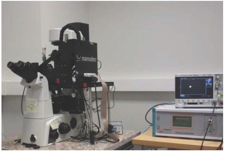
The equipment has eight independent piezoelectrics, Dulcinea® control, WSxM software, a fluid cell and an XY positioner. It allows AFM images to be taken in contact, jumping, lithographic and dynamic (amplitude and frequency modulation) modes; spectroscopic measurements in 2 (FZ), 3 (3D) and 4 (general spectroscopy imaging, including force volume) dimensions; and long-distance measurements with retrace and plane scan methods. It also includes automatic drift correction. Moreover, it has a liquid exchange system which makes it possible to control the physical and chemical conditions of the medium in which the specimens are held. The equipment is connected to an inverted optical microscope, which allows the sensor element to be positioned on any point of interest in the specimen.
Nanotribology, imaging, bioimaging, biophysics, interaction between individual molecules, surface topography, cell mechanics, cell-material interaction, mechanobiological characterisation of samples.
The AFM with fluid cell makes it possible to work in conditions where cells are not under stress. In addition, it is possible to control the conditions of the medium in which the cells are held, in order to observe the changes that occur in response to chemical or physical stress.
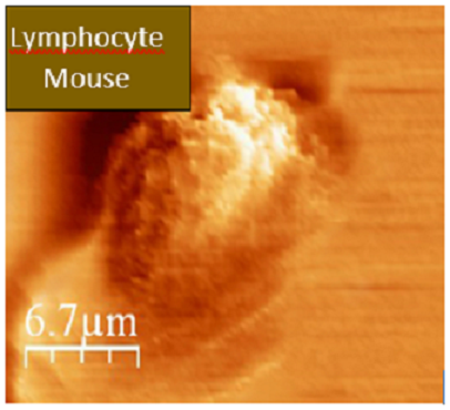
With this device, the mechanical properties of living biological entities can be imaged and studied in different media and under controlled conditions. Images of cells subjected to different types of chemical or physical stress can also be taken. As it is capable of achieving molecular resolution, the surfaces of materials can be seen at nanometric scales.
Affinity microscopy, single-cell force spectroscopy (SCFS) and single-molecule force spectroscopy (SMFS).


